


3D Printed Tracheoesophageal Fistula and Oesophagus Atresia
Clinical History
A 32-year-old female G3P0 (gravida 3, para 0‘ – i.e., has had two pregnancies,
with neither of the embyros surviving to a gestational age of 24 weeks) presents in preterm labour at 25 weeks
gestation. The GP had noted an increased fundal height at 30cm one week prior, but the mother had refused prenatal
testing or ultrasound, and was lost to follow up. She delivered a live born male baby. Examination of the baby noted
polydactyly, imperforate anus, excessive drooling, and a loud pan-systolic murmur. A single umbilical artery was
noted in the umbilical cord. The baby had difficulty feeding with increasing respiratory distress. The baby died 2
days later from aspiration pneumonia.
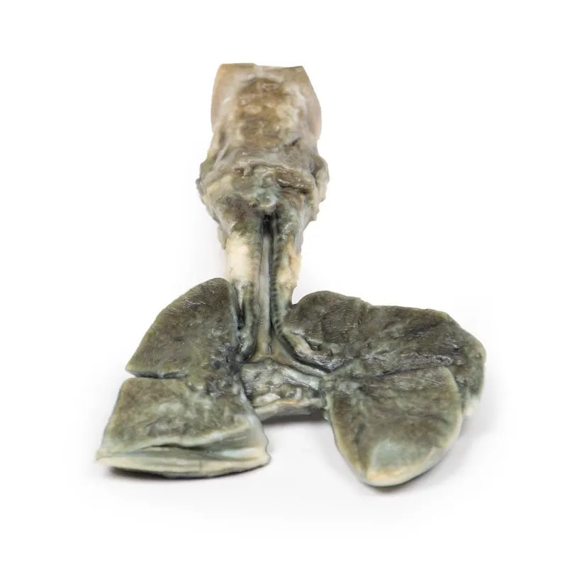
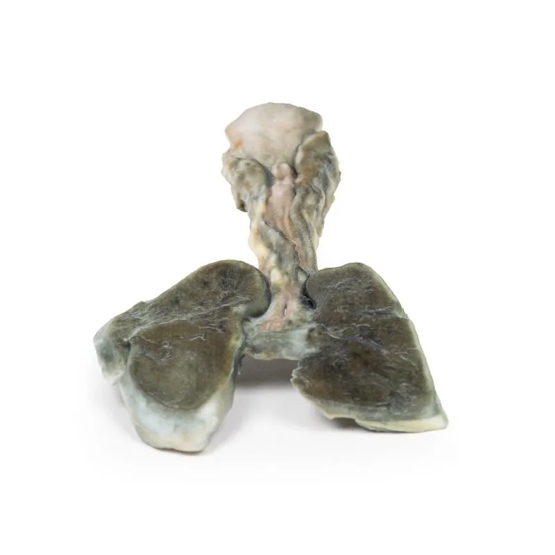
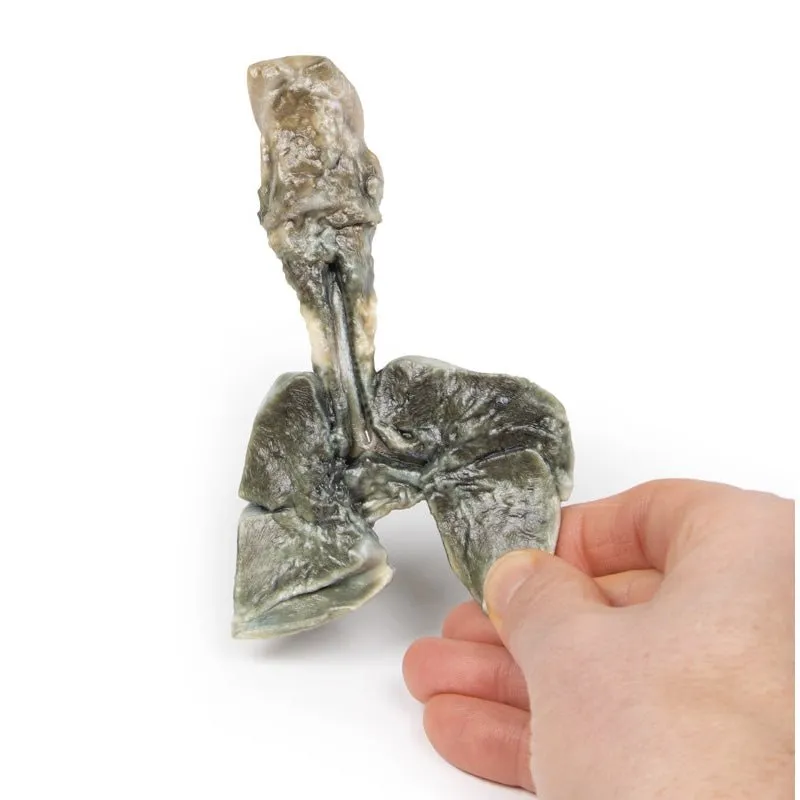
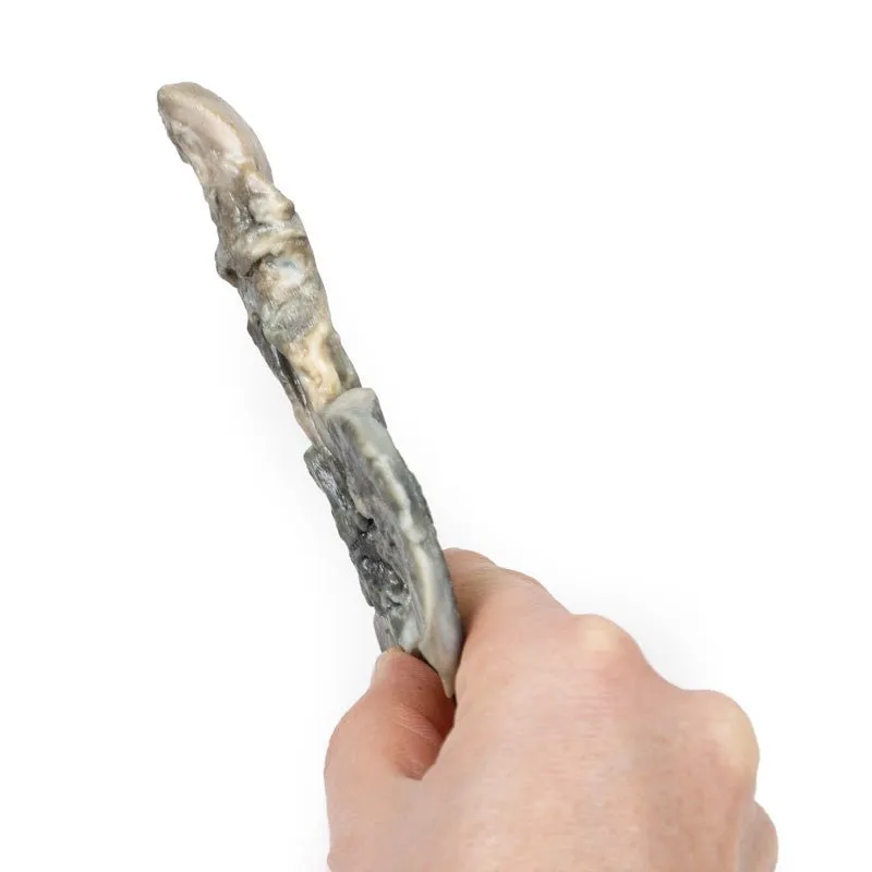
Pathology/Specimen Details
The specimen comprises the tongue, larynx, trachea, bronchi, both
lungs and oesophagus of the foetus. The trachea and bronchi have been divided in the midline. A fistula is present
just above the bifurcation at a communicating fistula can be seen connecting the distal oesophagus to the trachea
(arrow). This is an example of a Type C Tracheoesophageal Fistula (oesophageal atresia with distal tracheoesophageal
fistula). It is difficult to discern if the oesophagus ends as a blind pouch at the lower extent of the specimen.
Further Information
Tracheoesophageal Fistula (TEF) is a common congenital abnormality occurring
in about 1 in 4000 live births. TEF usually occurs with oesophageal atresia (sometimes abbreviated to EA, reflecting
the US spelling of ‘esophagus’). TEF are classified according to their anatomical configuration. Type C is the most
common configuration; as described above, in which oesophageal atresia with distal tracheoesophageal fistula making
up 86% of cases. TEF occurs without oesophageal atresia in only 4% of cases, Type E.
TEF and oesophageal atresia
are caused by defective lateral septation of the foregut into the oesophagus and trachea. It is believed that a
defect in epithelial-mesenchymal interactions causes failed branching of a lung bud branch which becomes the fistula
tract. It is associated with VACTERL (vertebral defects, anal atresia, cardiac defects, TEF, renal anomalies, and
limb abnormalities) or CHARGE syndrome (Coloboma, Heart defects, Atresia choanae, Growth retardation, Genital
abnormalities, and Ear abnormalities).
Oesophageal atresia can be seen on prenatal ultrasound as polyhydramnios, absent/collapsed stomach, and proximal
oesophageal pouch dilation. EA with TEF can be more difficult to see on ultrasound as fistula allows fluid flow into
the stomach. Polyhydramnios occurs in one third of cases of EA with distal TEF. Postnatal symptoms vary on the
configuration of the fistula. These include excessive drooling, respiratory distress, difficulty feeding and
choking. Reflux of gastric contents can lead to aspiration pneumonia as in this case.
Diagnosis can be made by
failing to pass a nasogastric tube into the stomach along with X-ray imaging. Fluoroscopy with contrast can be used
for more indeterminate cases. For milder cases diagnosis may be made later with endoscopic investigation. Treatment
involves surgical correction of the defects. Prognosis is usually good. However, cases with associated chromosomal,
prematurity and cardiac defects are at increased risk of death.
GTSimulators by Global Technologies
Erler Zimmer Authorized Dealer
")
![Miral Linen Table Cloth [Pale terracotta]](https://www.phototops.shop/image/miral-linen-table-cloth-pale-terracotta_oJH0jK_300x.webp "Miral Linen Table Cloth [Pale terracotta]")


